This web page was produced as an assignment for Genetics 677, an undergraduate course at UW-Madison
Protein Sequence
The RB1 Protein is 928 amino acids long. Protein sequence can be viewed by clicking on the link below.
FASTA Protein Sequence
Protein Function
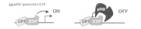
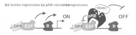
In addition to inhibiting transcription factor E2F, the Rb1 protein also attracts Histone deacetylase protein to surrounding chromatin, further suppressing DNA synthesis.(11)
In the figures below, it can be seen how Rb1 interacts with both E2F and histone deacetylase to inhibit DNA synthesis.
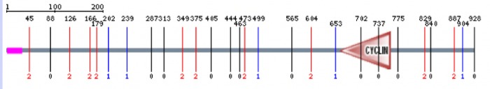
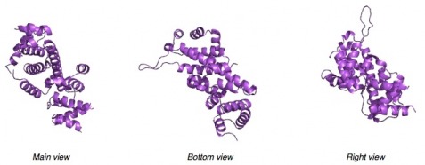
RB1 Protein Domains
RB1 has three known protein domains. The N-terminus, the C-terminus, and the A/B pocket.(11)
The N-terminus is not structurally well defined, but it is still known to be an important essential part of the protein because deletion mutants have been found in retinoblastomas. The N-terminus appears to interact with the A/B pocket. It is believed to promote an active Rb1 protein conformation. (11)
The C-terminus contains the cyclin binding motif. This domain is important for phosphorylation of the Rb1 protein, which is a control point for the activation or deactivation of the protein. The C-terminus is believed to interact with the A/B domain as a flexible regulatory arm. Cyclins, along with additional kinases form the maturation promoting factor (MPF). The MPF stimulates the entry into the mitotic and meiotic cell cycles. MPF is specifically involved in the control at the G1/S transition and the G2/Mitosis transition. (11)
The A/b pocket is the domain were many of the deleterious, deacivating mutations occur. The A/B pocket forms a repressor motif. The A portion forms a supportive scaffold for the B domain which ensures the proper folding and stability of the protein. (11)
Below is a figure of the protein with the cyclin domain evident and the N-terminus domain in purple on the left side of the protein. For unknown reasons, the figure didn't display and label the A/B domain (10)
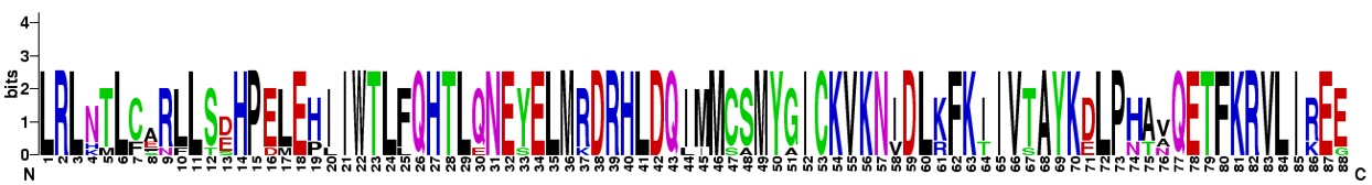
Multiple Sequence Alignment of the Cyclin Domain in RB1 Homologs
The cyclin domain in the human RB1 protein resides from amino acid 660 to amino acid 761. A multiple sequence alignment was generated using Web Logo and comparing the homologous cyclin domain between the RB1 homologs present in the chimpanzee, dog, norway rat, mouse, cattle, chicken, and zebra fish. The below image can be seen in full by downloading the picture.

| rb1weblogo.jpg | |
| File Size: | 88 kb |
| File Type: | jpg |
A sequence logo is a graphical representation of an aligned set of binding sites. Height of letters at each position represent frequencies and the total height at each position represents sequence conservation. As can be seen in the figure, the majority of the amino acid residues are conserved throughout the homologous cyclin domain. (9)
It was not that surprising that there was a high degree of conservation between the homologs, as the cyclin domain is essential for the proper function of the RB1 protein.
References
References
1. GeneBee http://www.genebee.msu.su/
2. ClustalW http://www.ebi.ac.uk/Tools/clustalw2/index.html
3. NCBI http://www.ncbi.nlm.nih.gov/
4. Homologene http://www.ncbi.nlm.nih.gov/homologene/
5. MOTIF http://motif.genome.jp/
6.Knudson 1971, Proc Acad Nat Sci USA 68:820
7. Figure 1, Retinoblastoma: the disease, gene and protein provide critical leads to understand cancer. DiCiomma D. et al. seminars in CANCER BIOLOGY, Vol. 10, 2000: pp. 255–269
8. Figure 2: SMART, http://smart.embl-heidelberg.de/
9. Figure 3: WEBLOGO, http://weblogo.berkeley.edu/
10.Lohmann DR, Gallie BL (2004). "Retinoblastoma: revisiting the model prototype of inherited cancer.". American journal of medical genetics. Part C, Seminars in medical genetics 129 (1): 23–8.
11.Retinoblastoma: the disease, gene and protein provide critical leads to understand cancer. DiCiomma D. et al. seminars in CANCER BIOLOGY, Vol. 10, 2000: pp. 255–269
12. Figure 4: Pfam, http://pfam.sanger.ac.uk/
